Tendon Diagram / Achilles Tendon Pain Causes Diagnosis And Treatment : Tendon diagram of calf and knee.. Brings trunk forward, and aids expiration. 19 photos of the knee tendon anatomy diagram and name chart. Ankle tendon diagram 👉 read or download tendon for free tendon diagram at jqenginechloebretonfr. The tendons have 2 functions: Tendons transmit the mechanical force of muscle contraction to the bones.
Diagram of knee tendons and ligaments. One tendons inserts onto the forearm bone, the radius, and the second spreads out to join the fascia along the upper part of the forearm. Diagram of inside the body. The golgi tendon organ (gto) (also called golgi organ, tendon organ, neurotendinous organ or neurotendinous spindle) is a proprioceptive sensory receptor organ that senses changes in muscle tension. When the biceps contracts, it pulls the forearm up and rotates it outward.
Basic Hand And Wrist Anatomy Hand Institute Of Charleston from handinstituteofcharleston.com Diagram of knee tendons and ligaments. Related posts of diagram of shoulder muscles and tendons neck muscle anatomy mri. Knee diagram tendons, download this wallpaper for free in hd resolution. Brings trunk forward, and aids expiration. Find symptoms,causes and treatments of joint disorders.for your health. Neck muscle anatomy mri 12 photos of the neck muscle anatomy mri neck muscle anatomy images, neck muscle anatomy pictures, neck muscle anatomy posterior, neck muscle anatomy ultrasound, neck muscles anatomy radiology, human muscles, neck muscle anatomy images, neck muscle anatomy pictures, neck muscle anatomy. Diagram of tendons in hand stock illustration. This diagram with labels depicts and explains the details of.
Tendons are similar to ligaments;
Here you can see the tendons that extend down the top of your foot toward your toes, allowing you to curl your toes upward if need be. Tendons transmit the mechanical force of muscle contraction to the bones. Foot anatomy diagram, foot joint diagram, foot sprain diagram, foot tendons and ligaments pain, leg tendon diagram. Tendon, tissue that attaches a muscle to other body parts, usually bones. Brings trunk forward, and aids expiration. Concertina tibial tendon diagram, generally known as dannert tibial tendon diagram is a form of barbed or razor tibial tendon diagram that is definitely fashioned in substantial coils which might be expanded similar to a concertina. Diagram of the ankle bones. Related posts of diagram of shoulder muscles and tendons neck muscle anatomy mri. Diagram depicting the bones, ligaments and muscles throughout the hand and fingers. Anatomy diagrams of shoulder, arm, elbow, forearm, wrist and hand. Tendon diagram of calf and knee. Tendon diagram of the knee. A change in shape of a muscle (the stimulus) causes the muscle to readjust its shape (the response) and maintain your posture.
The achilles tendon is also called the calcaneal tendon. The insertions of the tibialis posterior tendon are illustrated. The tendons have 2 functions: This forearm muscle is responsible for extending all of the fingers of the hand except the thumb. 1 article features images from this case.
Ligament Vs Tendon What S The Difference from i0.wp.com Concertina tibial tendon diagram, generally known as dannert tibial tendon diagram is a form of barbed or razor tibial tendon diagram that is definitely fashioned in substantial coils which might be expanded similar to a concertina. Learn vocabulary, terms and more with flashcards, games and other study tools. One tendons inserts onto the forearm bone, the radius, and the second spreads out to join the fascia along the upper part of the forearm. Tendon diagram of the knee. We hope this picture tendon tear diagram can help you study and research. Neck muscle anatomy mri 12 photos of the neck muscle anatomy mri neck muscle anatomy images, neck muscle anatomy pictures, neck muscle anatomy posterior, neck muscle anatomy ultrasound, neck muscles anatomy radiology, human muscles, neck muscle anatomy images, neck muscle anatomy pictures, neck muscle anatomy. Quadriceps tendon rupture is usually associated with forced flexion of the knee or a direct blow, although spontaneous ruptures are reported. If you would like to learn all the parts of the foot structure, you have come to the right place.
Anatomy diagrams of shoulder, arm, elbow, forearm, wrist and hand.
This diagram with labels depicts and explains the details of. Learn vocabulary, terms, and more with flashcards, games, and other study tools. Tendons are similar to ligaments; Cyst on the lower part of the diagram. Foot anatomy diagram, foot joint diagram, foot sprain diagram, foot tendons and ligaments pain, leg tendon diagram. When the muscles tighten (contract) arguably, the most important tendon is the achilles tendon, which allows the calf muscles to move. Concertina tibial tendon diagram, generally known as dannert tibial tendon diagram is a form of barbed or razor tibial tendon diagram that is definitely fashioned in substantial coils which might be expanded similar to a concertina. Learn about the anatomy and physiology of tendons. Also allows the action of raising up onto toes. Allows the action of raising the foot. Diagram of the ankle bones. Feet human anatomy bones tendons ligaments and more. Allows the foot to be turned inward and also supports the arch of the foot.
Tendons are similar to ligaments; The insertions of the tibialis posterior tendon are illustrated. Allows the action of raising the foot. The muscle belly then crosses the entire upper arm and separates into two tendons. Anatomy diagrams of shoulder, arm, elbow, forearm, wrist and hand.
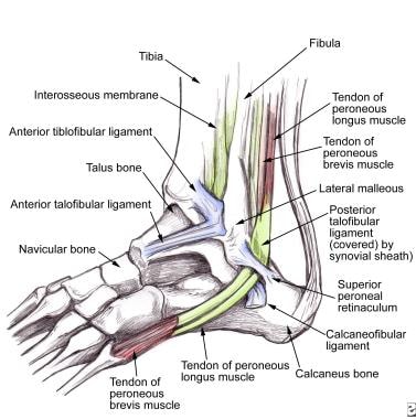
Peroneal Tendon Syndromes Practice Essentials Epidemiology Functional Anatomy from img.medscapestatic.com Diagram depicting the bones, ligaments and muscles throughout the hand and fingers. This forearm muscle is responsible for extending all of the fingers of the hand except the thumb. The golgi tendon organ (gto) (also called golgi organ, tendon organ, neurotendinous organ or neurotendinous spindle) is a proprioceptive sensory receptor organ that senses changes in muscle tension. Concertina tibial tendon diagram, generally known as dannert tibial tendon diagram is a form of barbed or razor tibial tendon diagram that is definitely fashioned in substantial coils which might be expanded similar to a concertina. Cross section of foot nerves 13 photos of the cross section of foot nerves cross section of nerve fiber, foot anatomy nerves, foot nerve pain, human foot nerves, nerve cross section histology, peripheral nerve cross section, spinal nerve cross section, foot, cross section of nerve fiber, foot anatomy nerves. Pin on custom made orthotics. Tendons are found throughout the body, from the head and neck all the way down to the feet. 1 article features images from this case.
Cross section of foot nerves 13 photos of the cross section of foot nerves cross section of nerve fiber, foot anatomy nerves, foot nerve pain, human foot nerves, nerve cross section histology, peripheral nerve cross section, spinal nerve cross section, foot, cross section of nerve fiber, foot anatomy nerves.
Tendons are similar to ligaments; Diagram depicting the bones, ligaments and muscles throughout the hand and fingers. Anatomy diagrams of shoulder, arm, elbow, forearm, wrist and hand. Brings trunk forward, and aids expiration. 1 article features images from this case. The insertions of the tibialis posterior tendon are illustrated. For example, a tap to the tendon under the knee cap elicits (triggers) the knee jerk reflex. Tendons, located at each end of a muscle, attach muscle to bone. Foot anatomy diagram, foot joint diagram, foot sprain diagram, foot tendons and ligaments pain, leg tendon diagram. Also allows the action of raising up onto toes. Tendon, tissue that attaches a muscle to other body parts, usually bones. When the biceps contracts, it pulls the forearm up and rotates it outward. Diagram of tendons in hand stock illustration.


Posting Komentar
Posting Komentar